Dental X-rays are an important part of routine dental care because they reveal problems that cannot be seen during a visual exam. Many people wonder whether dental X-rays are safe or necessary at every visit. Modern digital dental X-ray systems use very low radiation and give dentists a clear view of your teeth, gums, and jaw, helping detect issues early and accurately.
These images show cavities, hidden decay, infections, impacted teeth, and bone changes long before symptoms appear. Early detection makes treatment simpler, less invasive, and more affordable. Understanding what dental X-rays show and why dentists recommend them helps you make informed decisions about your oral health.
This guide explains everything you need to know about dental X-rays in clear and simple terms.
Table of Contents
ToggleWhat This Guide Covers
- What dental X-rays are and how they work
- How the test is performed using different types of X-rays
- Why dental exams and X-rays are an essential part of preventive care
- What dental radiographs can detect, including early decay and bone loss
- Different types of dental X-rays and when each one is used
- Risks, safety, and are dental X-rays are dangerous are concerns
- How many dental X-rays are safe, and when to avoid them
What Are Dental X-rays?
Dental X-rays, also called dental radiographs, are images used by your dentist to examine your teeth, gums, and jaw. A digital dental X-ray uses low levels of radiation to capture detailed images inside your mouth.
These images help detect issues such as:
- Cavities and tooth decay
- Impacted or missing teeth
- Bone loss in the jaw
- Infections or abscesses
In a digital dental X-ray process, X-rays pass through soft tissues and are absorbed by denser structures, such as teeth and fillings.
- Dense materials such as metal fillings appear white.
- Air spaces appear black.
- Teeth, tissues, and fluids appear in shades of gray.
Most modern dental clinics now use digital X-ray technology, which offers clearer images, less radiation, and faster results than traditional film X-rays.
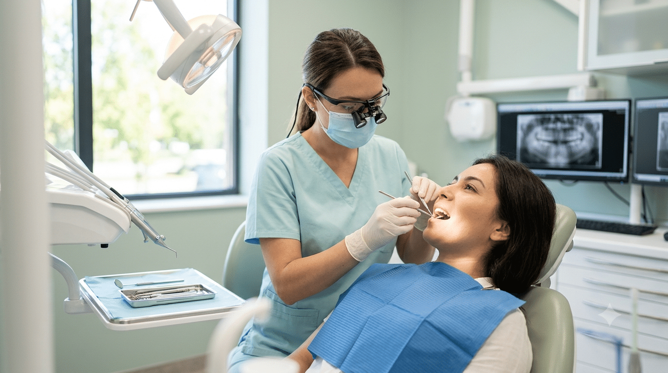
How the Test is Performed
A dental X-ray is done in the dentist’s office using different imaging methods. Many clinics use digital X-ray dental systems to reduce radiation and improve image quality. The process typically follows these steps:
Step 1: The dentist reviews your needs.
They decide which type of X-ray is required based on symptoms, treatment planning, or routine evaluation.
Step 2: The dental team positions you correctly.
You may sit, stand, or bite on a tab depending on the X-ray type.
Step 3: A protective apron is placed on you.
This helps limit unnecessary radiation exposure.
Step 4: The X-ray device is positioned.
Different devices are used for specific images:
- Bitewing: You bite on a tab to capture the crowns of upper and lower teeth.
- Periapical X-ray: Focuses on one or two full teeth from crown to root.
- Palatal or Occlusal X-ray: Shows all upper or lower teeth in one image using a film placed on the biting surface.
- Panoramic X-ray: A rotating machine captures the entire mouth, jaws, and surrounding areas.
- Cephalometric X-ray: Captures a side view of the face and jaw for orthodontic and airway evaluation.
Step 5: The image is captured within seconds.
Digital systems send the image instantly to a computer.
Step 6: 3D imaging is used if needed.
A 3D dental X-ray (Cone Beam CT) may be taken to create a complete 3D picture of the jaw, often before dental implant surgeries.
Step 7: The dentist reviews the results.
They check for decay, bone issues, tooth alignment, and other concerns based on the selected X-ray type.
Why the Test is Performed
Dental exams and X-rays are done to monitor your oral health and detect problems early. Many dentists take X-rays once a year, but they may be needed more often if your dentist is evaluating a specific issue or planning a treatment.
How often you need dental exams and X-rays depends on:
- Age
- Current oral health
- Symptoms of oral disease
- History of gum disease or tooth decay
If you are a new patient, your dentist may take X-rays to understand your dental history, especially if no previous records are available. These images illustrate what dental X-rays reveal, such as cavities, infections, and jawbone loss.
Children may need X-rays more often because dentists must track the growth of adult teeth. This helps determine whether baby teeth should be removed to prevent crowding or to prevent teeth from growing in the wrong position.
What Can Dental Radiographs Detect?
Dental X-rays show many conditions that may not be visible during a regular exam. A bad teeth X-ray can reveal:
- Cavities, including decay between teeth
- Decay under fillings
- Bone loss in jaw X-ray images
- Impacted or unerupted teeth
- Abscesses or infections
- Cysts and some tumors
Dentists also use digital dental X-rays to evaluate whether you are a candidate for implants, braces, or dentures. They help track healing after treatments such as root canals or bone grafts.
What Abnormal Results Mean
Abnormal X-ray findings may include:
- The number, size, and position of teeth
- Fully or partially impacted teeth
- Severity of tooth decay
- Bone damage from gum disease
- Abscessed teeth
- Fractured jaw concerns
- Bite alignment issues
- Other structural problems in the teeth or jaw bones
Types of Dental X-rays
There are two main types of dental X-rays based on where the film or sensor is placed.
Intraoral X-rays
The sensor is placed inside your mouth and shows detailed images of your teeth and gums.
- Bitewing X-ray: Shows cavities between teeth and below the gumline.
- Periapical X-ray: Detects gum disease, bone loss, and cavities near the tooth roots.
- Occlusal X-ray: Reveals issues under the tongue or on the roof of the mouth, including impacted teeth or jaw fractures.
Extraoral X-rays
The sensor stays outside your mouth and captures larger structures.
- Panoramic X-ray: Shows the entire mouth, including teeth, jaws, nerves, sinuses, and bone.
- Cephalometric X-ray: Provides a side view of the head for orthodontic planning.
- Cone Beam CT Scan: Creates 3D images of teeth, jaws, nerves, and sinuses, often used for implant planning.
Risks of Dental X-rays
Are dental X-rays safe? Dental X-rays give off very low radiation, and modern digital dental X-ray systems reduce exposure even further. Most patients, including children, receive only minimal radiation during routine imaging.
Your dentist limits exposure by using safety measures such as:
- A lead apron to protect the chest, abdomen, and pelvic area
- A thyroid collar for people with thyroid concerns, children, and women of childbearing age
These protective tools help prevent unnecessary contact with radiation.
Although the risk is low, no one should receive more radiation than necessary. Dentists follow strict guidelines and take X-rays only when needed for diagnosis or treatment.
Pregnancy remains an important exception. Are dental X-rays safe during pregnancy? Women who are pregnant or think they may be pregnant should avoid dental X-rays unless the situation is urgent. Radiation is not considered safe for a developing fetus, so it is important to tell your dentist if pregnancy is possible.
The radiation from dental X-rays is extremely small, and the concern that dental X-rays are dangerous is rarely a concern when proper precautions are used. Most risks decrease further when clinics use digital X-ray dental systems.
After Dental X-rays and Tips
Once the images are taken, your dentist reviews the digital dental X-ray results to detect cavities, infections, bone loss, and other abnormalities. If your teeth are being cleaned by a hygienist, the dentist may discuss the X-ray findings after the cleaning unless an urgent issue appears on the images.
If your dentist sees problems such as decay or early signs of gum disease, they will explain the condition and recommend treatment. If your X-rays show no concerns, continue your current oral care routine and follow your dentist’s advice.
Regular dental exams and X-rays are important for maintaining good oral health. Even if your checkup looks perfect, skipping future X-rays is not recommended, as they help detect hidden issues early.
How often you need X-rays depends on your age, oral health, and risk factors. Many patients need them every one to two years, while others may need them more often if they have a history of decay or gum disease.
To protect your oral health:
- Keep all scheduled dental visits.
- Report any pain, swelling, or changes in your mouth.
- Maintain daily brushing and flossing.
Lincolnwood Family Dental: Safe, Modern, and Caring Approach
Lincolnwood Family Dental is committed to providing exceptional care in a comfortable and friendly setting. The team focuses on helping every patient achieve a healthy, confident smile using modern tools, including digital X-ray systems for safe, accurate diagnosis.
The clinic offers a wide range of services, including:
- Preventive dentistry and general dentistry
- Kids dentistry and emergency care
- Dental implants and wisdom teeth removal
- Invisalign and other orthodontic solutions
- Crowns, dentures, partials, veneers, and cosmetic dentistry
These services support patients of all ages, whether they need routine exams or advanced treatment. The team explains each procedure clearly and uses digital imaging to help patients understand their oral health.
Lincolnwood Family Dental also supports patients by simplifying insurance use. They are in-network with all PPO providers, including Aetna, Blue Cross Blue Shield, Careington, Cigna, Delta Dental, GEHA, Guardian, Humana, MetLife, Principal, Unicare, United Concordia, and United Healthcare. The staff helps patients understand and maximize their benefits.
Frequently Asked Questions
How long does a dental X-ray take?
Most digital dental X-rays take only a few seconds to capture, and the entire process usually finishes within a few minutes.
Can I eat or drink before a dental X-ray?
Yes. There are no food or drink restrictions before standard dental X-rays.
Do dental X-rays hurt?
No. You may feel slight pressure from the sensor or tab inside your mouth, but the process is not painful.
Can dental X-rays detect sinus problems?
Some X-rays, especially panoramic or 3D images, may show parts of the sinus area and help identify related issues.
Do I need to remove jewelry for a dental X-ray?
You may need to remove earrings, facial piercings, or glasses if they interfere with the image.